Intriguing connection found between serotonin and fertility

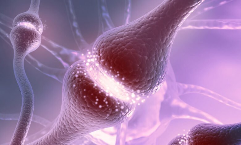
What role does the neurotransmitter serotonin play in fertility? A recent study by scientists from Nagoya University in Japan has uncovered a link between serotonin neurons, glucose availability, and reproductive health. Their findings suggest that serotonin neurons in the brain play a significant role in maintaining reproductive functions by sensing glucose levels and enhancing the release of reproductive hormones. The research was published in Scientific Reports.
Depression is linked to dysfunction in central serotonergic neurons and is known to correlate with both reproductive and metabolic disorders. Serotonin reuptake inhibitors, common treatments for depression, highlight the importance of serotonergic signaling in these processes.
However, the specific role of serotonergic neurons in coordinating reproduction and glucose metabolism remained unclear. The researchers aimed to uncover how serotonergic neurons in the brain might sense glucose levels and regulate reproductive functions. Understanding this connection could pave the way for new therapeutic approaches to treat reproductive disorders in patients with depression.
To investigate this, the researchers employed both female rats and goats, focusing on the dorsal raphe nucleus and the arcuate nucleus of the hypothalamus, areas rich in serotonergic neurons and key regulators of reproductive hormones. They used a combination of genetic, pharmacological, and physiological techniques to unravel the connections between these neurons, glucose sensing, and reproductive hormone release.
In rats, the team used Kiss1-tdTomato heterozygous rats, which have a gene marker that allows for the identification of kisspeptin neurons, crucial for regulating reproduction. They conducted RNA sequencing to identify the types of serotonin receptors present in these neurons. The analysis revealed that the serotonin-2C receptor (5HT2CR), an excitatory receptor, was significantly expressed in the arcuate nucleus’ kisspeptin neurons.
One of the primary discoveries is the significant expression of the serotonin-2C receptor (5HT2CR) in kisspeptin neurons within the arcuate nucleus of the hypothalamus. Kisspeptin neurons are crucial regulators of reproduction, responsible for generating pulses of gonadotropin-releasing hormone (GnRH), which in turn stimulates the release of luteinizing hormone (LH) necessary for reproductive processes.
The RNA sequencing and double in situ hybridization techniques used in this study confirmed that nearly half of the kisspeptin neurons expressed 5HT2CR. This finding suggests that these neurons are direct targets for serotonergic signaling, linking serotonin levels to reproductive function.
In further experiments, the researchers demonstrated that enhancing serotonergic activity in the mediobasal hypothalamus with fluoxetine, a serotonin reuptake inhibitor, could counteract the suppression of LH pulses induced by a glucoprivic state (low glucose availability) in female rats.
Normally, conditions of low glucose, induced experimentally by the administration of 2-deoxy-D-glucose (2DG), suppress the release of LH, thereby inhibiting reproductive functions. However, fluoxetine administration restored LH pulse frequency, indicating that increased serotonin levels can mitigate the adverse effects of low glucose on reproductive hormone release.
The researchers also showed that direct glucose infusion into the dorsal raphe nucleus increased serotonin release in the mediobasal hypothalamus. This intervention also restored the frequency of LH pulses suppressed by 2DG-induced glucoprivation. These results suggest that serotonergic neurons in the dorsal raphe can sense glucose levels and adjust reproductive hormone release accordingly, highlighting the dual role of these neurons in managing both glucose metabolism and reproductive function.
To validate these findings in another mammalian model, the researchers conducted electrophysiological recordings in goats. They discovered that central administration of serotonin or a 5HT2CR agonist stimulated the activity of the GnRH pulse generator, leading to increased LH release. Conversely, when a 5HT2CR antagonist was administered, it blocked the serotonin-induced stimulation of GnRH pulses, further confirming the pivotal role of the serotonin-2C receptor in this regulatory process.
The findings underscore the importance of serotonergic signaling in the brain’s ability to integrate information about glucose availability and modulate reproductive functions. The study provides evidence that serotonergic neurons, through their ability to sense glucose and interact with kisspeptin neurons via 5HT2CR, play a crucial role in maintaining reproductive health, particularly in the face of metabolic challenges.
Animal models, such as the female rats and goats used in this study, provide valuable insights into biological processes that are often difficult to study directly in humans. Rats and goats, while different from humans, share fundamental aspects of their endocrine and nervous systems. The findings in these animals can thus provide a basis for understanding similar processes in humans.
But human physiology, behavior, and disease pathology are influenced by a myriad of genetic, environmental, and social factors that are not fully replicated in animal models. For example, while rats and goats can provide insights into basic physiological processes, they do not capture the full complexity of human reproductive and metabolic disorders, which can be affected by a wide range of factors including diet, lifestyle, and psychological stressors.
While animal models are crucial for initial discoveries, further research involving human subjects is necessary to validate and translate these findings into clinical practice.
The study, “Raphe glucose-sensing serotonergic neurons stimulate KNDy neurons to enhance LH pulses via 5HT2CR: rat and goat studies,” was authored by Sho Nakamura, Takuya Sasaki, Yoshihisa Uenoyama, Naoko Inoue, Marina Nakanishi, Koki Yamada, Ai Morishima, Reika Suzumura, Yuri Kitagawa, Yasuhiro Morita, Satoshi Ohkura, and Hiroko Tsukamura.
Source link


